Filters
Host (768597)
Bovine (1090)Canine (20)Cat (408)Chicken (1642)Cod (2)Cow (333)Crab (15)Dog (524)Dolphin (2)Duck (13)E Coli (239129)Equine (7)Feline (1864)Ferret (306)Fish (125)Frog (55)Goat (36847)Guinea Pig (752)Hamster (1376)Horse (903)Insect (2053)Mammalian (512)Mice (6)Monkey (601)Mouse (96266)Pig (197)Porcine (70)Rabbit (358709)Rat (11723)Ray (55)Salamander (4)Salmon (15)Shark (3)Sheep (4247)Snake (4)Swine (301)Turkey (57)Whale (3)Yeast (5336)Zebrafish (3022)Isotype (156643)
IgA (13624)IgA1 (941)IgA2 (318)IgD (1949)IgE (5594)IgG (87187)IgG1 (16733)IgG2 (1329)IgG3 (2719)IgG4 (1689)IgM (22029)IgY (2531)Label (239340)
AF488 (2465)AF594 (662)AF647 (2324)ALEXA (11546)ALEXA FLUOR 350 (255)ALEXA FLUOR 405 (260)ALEXA FLUOR 488 (672)ALEXA FLUOR 532 (260)ALEXA FLUOR 555 (274)ALEXA FLUOR 568 (253)ALEXA FLUOR 594 (299)ALEXA FLUOR 633 (262)ALEXA FLUOR 647 (607)ALEXA FLUOR 660 (252)ALEXA FLUOR 680 (422)ALEXA FLUOR 700 (2)ALEXA FLUOR 750 (414)ALEXA FLUOR 790 (215)Alkaline Phosphatase (825)Allophycocyanin (32)ALP (387)AMCA (80)AP (1160)APC (15217)APC C750 (13)Apc Cy7 (1248)ATTO 390 (3)ATTO 488 (6)ATTO 550 (1)ATTO 594 (5)ATTO 647N (4)AVI (53)Beads (225)Beta Gal (2)BgG (1)BIMA (6)Biotin (27817)Biotinylated (1810)Blue (708)BSA (878)BTG (46)C Terminal (688)CF Blue (19)Colloidal (22)Conjugated (29246)Cy (163)Cy3 (390)Cy5 (2041)Cy5 5 (2469)Cy5 PE (1)Cy7 (3638)Dual (170)DY549 (3)DY649 (3)Dye (1)DyLight (1430)DyLight 405 (7)DyLight 488 (216)DyLight 549 (17)DyLight 594 (84)DyLight 649 (3)DyLight 650 (35)DyLight 680 (17)DyLight 800 (21)Fam (5)Fc Tag (8)FITC (30165)Flag (208)Fluorescent (146)GFP (563)GFP Tag (164)Glucose Oxidase (59)Gold (511)Green (580)GST (711)GST Tag (315)HA Tag (430)His (619)His Tag (492)Horseradish (550)HRP (12960)HSA (249)iFluor (16571)Isoform b (31)KLH (88)Luciferase (105)Magnetic (254)MBP (338)MBP Tag (87)Myc Tag (398)OC 515 (1)Orange (78)OVA (104)Pacific Blue (213)Particle (64)PE (33571)PerCP (8438)Peroxidase (1380)POD (11)Poly Hrp (92)Poly Hrp40 (13)Poly Hrp80 (3)Puro (32)Red (2440)RFP Tag (63)Rhodamine (607)RPE (910)S Tag (194)SCF (184)SPRD (351)Streptavidin (55)SureLight (77)T7 Tag (97)Tag (4710)Texas (1249)Texas Red (1231)Triple (10)TRITC (1401)TRX tag (87)Unconjugated (2110)Unlabeled (218)Yellow (84)Pathogen (489613)
Adenovirus (8665)AIV (315)Bordetella (25035)Borrelia (18281)Candida (17817)Chikungunya (638)Chlamydia (17650)CMV (121394)Coronavirus (5948)Coxsackie (854)Dengue (2868)EBV (1510)Echovirus (215)Enterovirus (677)Hantavirus (254)HAV (905)HBV (2095)HHV (873)HIV (7865)hMPV (300)HSV (2356)HTLV (634)Influenza (22132)Isolate (1208)KSHV (396)Lentivirus (3755)Lineage (3025)Lysate (127759)Marek (93)Measles (1163)Parainfluenza (1681)Poliovirus (3030)Poxvirus (74)Rabies (1519)Reovirus (527)Retrovirus (1069)Rhinovirus (507)Rotavirus (5346)RSV (1781)Rubella (1070)SIV (277)Strain (67790)Vaccinia (7233)VZV (666)WNV (363)Species (2982223)
Alligator (10)Bovine (159546)Canine (120648)Cat (13082)Chicken (113771)Cod (1)Cow (2030)Dog (12745)Dolphin (21)Duck (9567)Equine (2004)Feline (996)Ferret (259)Fish (12797)Frog (1)Goat (90451)Guinea Pig (87888)Hamster (36959)Horse (41226)Human (955186)Insect (653)Lemur (119)Lizard (24)Monkey (110914)Mouse (470743)Pig (26204)Porcine (131703)Rabbit (127597)Rat (347841)Ray (442)Salmon (348)Seal (8)Shark (29)Sheep (104984)Snake (12)Swine (511)Toad (4)Turkey (244)Turtle (75)Whale (45)Zebrafish (535)Technique (5597646)
Activation (170393)Activity (10733)Affinity (44631)Agarose (2604)Aggregation (199)Antigen (135358)Apoptosis (27447)Array (2022)Blocking (71767)Blood (8528)Blot (10966)ChiP (815)Chromatin (6286)Colorimetric (9913)Control (80065)Culture (3218)Cytometry (5481)Depletion (54)DNA (172449)Dot (233)EIA (1039)Electron (6275)Electrophoresis (254)Elispot (1294)Enzymes (52671)Exosome (4280)Extract (1090)Fab (2230)FACS (43)FC (80929)Flow (6666)Fluorometric (1407)Formalin (97)Frozen (2671)Functional (708)Gel (2484)HTS (136)IF (12906)IHC (16566)Immunoassay (1589)Immunofluorescence (4119)Immunohistochemistry (72)Immunoprecipitation (68)intracellular (5602)IP (2840)iPSC (259)Isotype (8791)Lateral (1585)Lenti (319416)Light (37250)Microarray (47)MicroRNA (4834)Microscopy (52)miRNA (88044)Monoclonal (516109)Multi (3844)Multiplex (302)Negative (4261)PAGE (2520)Panel (1520)Paraffin (2587)PBS (20270)PCR (9)Peptide (276160)PerCP (13759)Polyclonal (2762994)Positive (6335)Precipitation (61)Premix (130)Primers (3467)Probe (2627)Profile (229)Pure (7808)Purification (15)Purified (78305)Real Time (3042)Resin (2955)Reverse (2435)RIA (460)RNAi (17)Rox (1022)RT PCR (6608)Sample (2667)SDS (1527)Section (2895)Separation (86)Sequencing (122)Shift (22)siRNA (319447)Standard (42468)Sterile (10170)Strip (1863)Taq (2)Tip (1176)Tissue (42812)Tube (3306)Vitro (3577)Vivo (981)WB (2515)Western Blot (10683)Tissue (2015946)
Adenocarcinoma (1075)Adipose (3459)Adrenal (657)Adult (4883)Amniotic (65)Animal (2447)Aorta (436)Appendix (89)Array (2022)Ascites (4377)Bile Duct (20)Bladder (1672)Blood (8528)Bone (27330)Brain (31189)Breast (10917)Calvaria (28)Carcinoma (13493)cDNA (58547)Cell (413805)Cellular (9357)Cerebellum (700)Cervix (232)Child (1)Choroid (19)Colon (3911)Connective (3601)Contaminant (3)Control (80065)Cord (661)Corpus (148)Cortex (698)Dendritic (1849)Diseased (265)Donor (1360)Duct (861)Duodenum (643)Embryo (425)Embryonic (4583)Endometrium (463)Endothelium (1424)Epidermis (166)Epithelium (4221)Esophagus (716)Exosome (4280)Eye (2033)Female (475)Frozen (2671)Gallbladder (155)Genital (5)Gland (3436)Granulocyte (8981)Heart (6850)Hela (413)Hippocampus (325)Histiocytic (74)Ileum (201)Insect (4880)Intestine (1944)Isolate (1208)Jejunum (175)Kidney (8075)Langerhans (283)Leukemia (21541)Liver (17340)Lobe (835)Lung (6064)Lymph (1208)Lymphatic (639)lymphocyte (22572)Lymphoma (12782)Lysate (127759)Lysosome (2813)Macrophage (31794)Male (1617)Malignant (1465)Mammary (1985)Mantle (1042)Marrow (2210)Mastocytoma (3)Matched (11710)Medulla (156)Melanoma (15522)Membrane (105772)Metastatic (3574)Mitochondrial (160319)Muscle (37419)Myeloma (748)Myocardium (11)Nerve (6398)Neuronal (17028)Node (1206)Normal (9486)Omentum (10)Ovarian (2509)Ovary (1172)Pair (47185)Pancreas (2843)Panel (1520)Penis (64)Peripheral (1912)Pharynx (122)Pituitary (5411)Placenta (4038)Prostate (9423)Proximal (318)Rectum (316)Region (202210)Retina (956)Salivary (3119)Sarcoma (6946)Section (2895)Serum (24880)Set (167654)Skeletal (13628)Skin (1879)Smooth (7577)Spinal (424)Spleen (2292)Stem (8892)Stomach (925)Stroma (49)Subcutaneous (47)Testis (15393)Thalamus (127)Thoracic (60)Throat (40)Thymus (2986)Thyroid (14121)Tongue (140)Total (10135)Trachea (227)Transformed (175)Tubule (48)Tumor (76921)Umbilical (208)Ureter (73)Urinary (2466)Uterine (303)Uterus (414)Human Plasminogen Activator, Urokinase Receptor (uPAR / PLAUR) Protein: Structure, Function, and Research Relevance
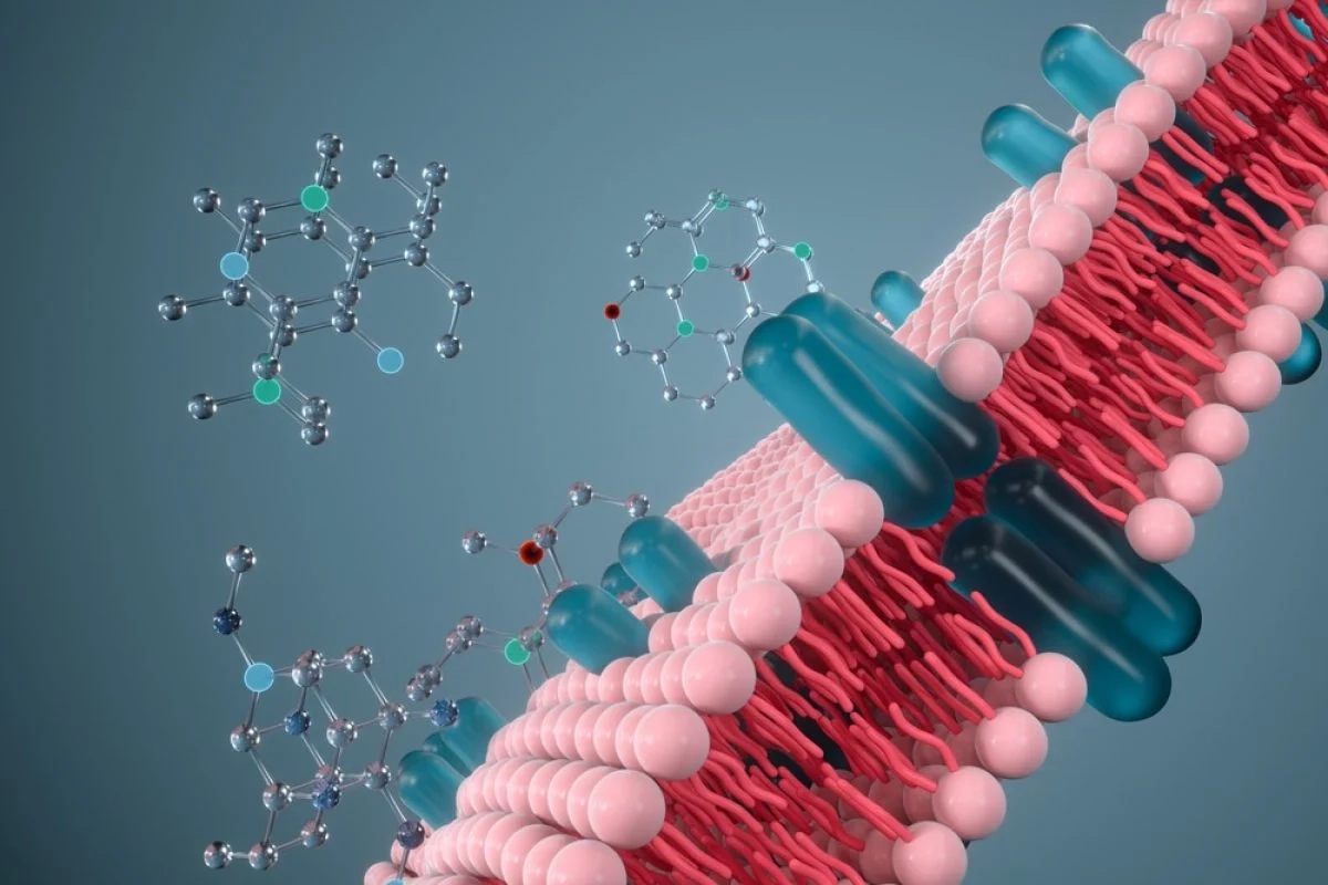
The urokinase plasminogen activator receptor (uPAR, also called PLAUR or CD87) is a crucial regulator of cell surface proteolysis and signaling. Anchored by a glycosylphosphatidylinositol (GPI) tail, uPAR localizes enzymatic activity of urokinase-type plasminogen activator (uPA), controlling extracellular matrix (ECM) degradation, wound healing, and tumor invasion.
Recombinant forms of the human uPAR protein are indispensable in research exploring metastasis, tissue regeneration, and biomarker discovery.
Genprice
Scientific Publications

Human Plasminogen Activator, Urokinase Receptor (uPAR / PLAUR) Protein: Structure, Function, and Research Relevance
Molecular Structure and Domain Organization
uPAR is composed of three Ly6/uPAR domains (D1–D3) that fold into a compact, “three-finger” architecture characteristic of the LU protein superfamily. These domains collectively form a high-affinity binding pocket for urokinase (uPA).
Unlike most receptors, uPAR lacks transmembrane and cytoplasmic segments, depending instead on lateral interactions with integrins and receptor tyrosine kinases (RTKs) to trigger downstream signaling cascades.
Schematic structure of uPAR showing the three-domain (D1–D3) organization and GPI-anchored membrane attachment. The diagram also illustrates urokinase binding and plasminogen activation localized at the cell surface.
Mechanisms of Action: From Proteolysis to Signaling
The uPA/uPAR system operates as a surface-bound catalytic unit:
- Step 1: uPAR captures pro-uPA or active uPA on the cell membrane.
- Step 2: uPA converts plasminogen to plasmin, which degrades ECM proteins.
- Step 3: The resulting proteolytic fragments and growth factors promote cell movement, tissue remodeling, and signaling.
Even beyond proteolysis, uPAR participates in non-enzymatic signaling by coupling with β1/β3 integrins, EGFR, and vitronectin, promoting cell adhesion, migration, and epithelial-mesenchymal transition (EMT). This makes it a key factor in both cancer progression and tissue repair.
Clinical Relevance and Disease Association
Cancer Invasion and Metastasis
Overexpression of uPAR has been documented in numerous cancers — breast, prostate, glioblastoma, and colorectal — correlating with tumor aggressiveness and metastatic potential. Imaging studies using uPAR-specific probes show how receptor density correlates with invasive front regions of tumors.
Immunofluorescence image showing uPAR localization in cancer cells. Co-staining with actin cytoskeleton (red) and uPAR (green) demonstrates enrichment at invasive protrusions, consistent with its role in matrix degradation and motility.
This dual role—anchoring enzymatic activity and coordinating migration—makes uPAR a potent biomarker and therapeutic target. Blocking uPAR–uPA interactions has shown promise in preclinical models by reducing invasion and angiogenesis.
Inflammation, Fibrosis, and suPAR as a Biomarker
The soluble form of uPAR (suPAR), released upon proteolytic cleavage, circulates in plasma and serves as a marker for immune activation and tissue injury. Elevated suPAR levels are linked to chronic inflammation, renal failure, cardiovascular disease, and fibrosis progression.
In nephrology, suPAR has emerged as a predictive biomarker for glomerular injury and podocyte dysfunction.
Experimental Applications of Recombinant Human uPAR
The availability of recombinant human uPAR proteins enables a variety of cutting-edge experimental approaches:
- Ligand-binding studies: Quantifying affinity between uPAR and uPA via SPR, ELISA, or fluorescence assays.
- Inhibitor screening: Testing peptides or antibodies designed to block the uPA–uPAR interface.
- Cell migration assays: Measuring the impact of uPAR overexpression or inhibition on motility.
- Structural and imaging studies: Using recombinant uPAR to resolve 3D structures or as a target for fluorescent tracers.
Researchers using GenPrice’s Human uPAR Protein can verify its suitability for binding and inhibition assays, ensuring biologically active conformation and purity above 95%.
Conclusion
The Human Plasminogen Activator, Urokinase Receptor (uPAR) is far more than a surface anchor for proteases—it’s a multifunctional signaling hub bridging the extracellular matrix, cell adhesion, and immune communication.
Studying uPAR using validated recombinant proteins, such as those offered by GenPrice, provides invaluable insights into molecular mechanisms underlying cancer, inflammation, and tissue regeneration.
🔗 Explore the product:
👉 Buy Human Plasminogen Activator, Urokinase Receptor (uPAR) Protein – GenPrice