Filters
Host (768597)
Bovine (1090)Canine (20)Cat (408)Chicken (1642)Cod (2)Cow (333)Crab (15)Dog (524)Dolphin (2)Duck (13)E Coli (239129)Equine (7)Feline (1864)Ferret (306)Fish (125)Frog (55)Goat (36847)Guinea Pig (752)Hamster (1376)Horse (903)Insect (2053)Mammalian (512)Mice (6)Monkey (601)Mouse (96266)Pig (197)Porcine (70)Rabbit (358709)Rat (11723)Ray (55)Salamander (4)Salmon (15)Shark (3)Sheep (4247)Snake (4)Swine (301)Turkey (57)Whale (3)Yeast (5336)Zebrafish (3022)Isotype (156643)
IgA (13624)IgA1 (941)IgA2 (318)IgD (1949)IgE (5594)IgG (87187)IgG1 (16733)IgG2 (1329)IgG3 (2719)IgG4 (1689)IgM (22029)IgY (2531)Label (239340)
AF488 (2465)AF594 (662)AF647 (2324)ALEXA (11546)ALEXA FLUOR 350 (255)ALEXA FLUOR 405 (260)ALEXA FLUOR 488 (672)ALEXA FLUOR 532 (260)ALEXA FLUOR 555 (274)ALEXA FLUOR 568 (253)ALEXA FLUOR 594 (299)ALEXA FLUOR 633 (262)ALEXA FLUOR 647 (607)ALEXA FLUOR 660 (252)ALEXA FLUOR 680 (422)ALEXA FLUOR 700 (2)ALEXA FLUOR 750 (414)ALEXA FLUOR 790 (215)Alkaline Phosphatase (825)Allophycocyanin (32)ALP (387)AMCA (80)AP (1160)APC (15217)APC C750 (13)Apc Cy7 (1248)ATTO 390 (3)ATTO 488 (6)ATTO 550 (1)ATTO 594 (5)ATTO 647N (4)AVI (53)Beads (225)Beta Gal (2)BgG (1)BIMA (6)Biotin (27817)Biotinylated (1810)Blue (708)BSA (878)BTG (46)C Terminal (688)CF Blue (19)Colloidal (22)Conjugated (29246)Cy (163)Cy3 (390)Cy5 (2041)Cy5 5 (2469)Cy5 PE (1)Cy7 (3638)Dual (170)DY549 (3)DY649 (3)Dye (1)DyLight (1430)DyLight 405 (7)DyLight 488 (216)DyLight 549 (17)DyLight 594 (84)DyLight 649 (3)DyLight 650 (35)DyLight 680 (17)DyLight 800 (21)Fam (5)Fc Tag (8)FITC (30165)Flag (208)Fluorescent (146)GFP (563)GFP Tag (164)Glucose Oxidase (59)Gold (511)Green (580)GST (711)GST Tag (315)HA Tag (430)His (619)His Tag (492)Horseradish (550)HRP (12960)HSA (249)iFluor (16571)Isoform b (31)KLH (88)Luciferase (105)Magnetic (254)MBP (338)MBP Tag (87)Myc Tag (398)OC 515 (1)Orange (78)OVA (104)Pacific Blue (213)Particle (64)PE (33571)PerCP (8438)Peroxidase (1380)POD (11)Poly Hrp (92)Poly Hrp40 (13)Poly Hrp80 (3)Puro (32)Red (2440)RFP Tag (63)Rhodamine (607)RPE (910)S Tag (194)SCF (184)SPRD (351)Streptavidin (55)SureLight (77)T7 Tag (97)Tag (4710)Texas (1249)Texas Red (1231)Triple (10)TRITC (1401)TRX tag (87)Unconjugated (2110)Unlabeled (218)Yellow (84)Pathogen (489613)
Adenovirus (8665)AIV (315)Bordetella (25035)Borrelia (18281)Candida (17817)Chikungunya (638)Chlamydia (17650)CMV (121394)Coronavirus (5948)Coxsackie (854)Dengue (2868)EBV (1510)Echovirus (215)Enterovirus (677)Hantavirus (254)HAV (905)HBV (2095)HHV (873)HIV (7865)hMPV (300)HSV (2356)HTLV (634)Influenza (22132)Isolate (1208)KSHV (396)Lentivirus (3755)Lineage (3025)Lysate (127759)Marek (93)Measles (1163)Parainfluenza (1681)Poliovirus (3030)Poxvirus (74)Rabies (1519)Reovirus (527)Retrovirus (1069)Rhinovirus (507)Rotavirus (5346)RSV (1781)Rubella (1070)SIV (277)Strain (67790)Vaccinia (7233)VZV (666)WNV (363)Species (2982223)
Alligator (10)Bovine (159546)Canine (120648)Cat (13082)Chicken (113771)Cod (1)Cow (2030)Dog (12745)Dolphin (21)Duck (9567)Equine (2004)Feline (996)Ferret (259)Fish (12797)Frog (1)Goat (90451)Guinea Pig (87888)Hamster (36959)Horse (41226)Human (955186)Insect (653)Lemur (119)Lizard (24)Monkey (110914)Mouse (470743)Pig (26204)Porcine (131703)Rabbit (127597)Rat (347841)Ray (442)Salmon (348)Seal (8)Shark (29)Sheep (104984)Snake (12)Swine (511)Toad (4)Turkey (244)Turtle (75)Whale (45)Zebrafish (535)Technique (5597646)
Activation (170393)Activity (10733)Affinity (44631)Agarose (2604)Aggregation (199)Antigen (135358)Apoptosis (27447)Array (2022)Blocking (71767)Blood (8528)Blot (10966)ChiP (815)Chromatin (6286)Colorimetric (9913)Control (80065)Culture (3218)Cytometry (5481)Depletion (54)DNA (172449)Dot (233)EIA (1039)Electron (6275)Electrophoresis (254)Elispot (1294)Enzymes (52671)Exosome (4280)Extract (1090)Fab (2230)FACS (43)FC (80929)Flow (6666)Fluorometric (1407)Formalin (97)Frozen (2671)Functional (708)Gel (2484)HTS (136)IF (12906)IHC (16566)Immunoassay (1589)Immunofluorescence (4119)Immunohistochemistry (72)Immunoprecipitation (68)intracellular (5602)IP (2840)iPSC (259)Isotype (8791)Lateral (1585)Lenti (319416)Light (37250)Microarray (47)MicroRNA (4834)Microscopy (52)miRNA (88044)Monoclonal (516109)Multi (3844)Multiplex (302)Negative (4261)PAGE (2520)Panel (1520)Paraffin (2587)PBS (20270)PCR (9)Peptide (276160)PerCP (13759)Polyclonal (2762994)Positive (6335)Precipitation (61)Premix (130)Primers (3467)Probe (2627)Profile (229)Pure (7808)Purification (15)Purified (78305)Real Time (3042)Resin (2955)Reverse (2435)RIA (460)RNAi (17)Rox (1022)RT PCR (6608)Sample (2667)SDS (1527)Section (2895)Separation (86)Sequencing (122)Shift (22)siRNA (319447)Standard (42468)Sterile (10170)Strip (1863)Taq (2)Tip (1176)Tissue (42812)Tube (3306)Vitro (3577)Vivo (981)WB (2515)Western Blot (10683)Tissue (2015946)
Adenocarcinoma (1075)Adipose (3459)Adrenal (657)Adult (4883)Amniotic (65)Animal (2447)Aorta (436)Appendix (89)Array (2022)Ascites (4377)Bile Duct (20)Bladder (1672)Blood (8528)Bone (27330)Brain (31189)Breast (10917)Calvaria (28)Carcinoma (13493)cDNA (58547)Cell (413805)Cellular (9357)Cerebellum (700)Cervix (232)Child (1)Choroid (19)Colon (3911)Connective (3601)Contaminant (3)Control (80065)Cord (661)Corpus (148)Cortex (698)Dendritic (1849)Diseased (265)Donor (1360)Duct (861)Duodenum (643)Embryo (425)Embryonic (4583)Endometrium (463)Endothelium (1424)Epidermis (166)Epithelium (4221)Esophagus (716)Exosome (4280)Eye (2033)Female (475)Frozen (2671)Gallbladder (155)Genital (5)Gland (3436)Granulocyte (8981)Heart (6850)Hela (413)Hippocampus (325)Histiocytic (74)Ileum (201)Insect (4880)Intestine (1944)Isolate (1208)Jejunum (175)Kidney (8075)Langerhans (283)Leukemia (21541)Liver (17340)Lobe (835)Lung (6064)Lymph (1208)Lymphatic (639)lymphocyte (22572)Lymphoma (12782)Lysate (127759)Lysosome (2813)Macrophage (31794)Male (1617)Malignant (1465)Mammary (1985)Mantle (1042)Marrow (2210)Mastocytoma (3)Matched (11710)Medulla (156)Melanoma (15522)Membrane (105772)Metastatic (3574)Mitochondrial (160319)Muscle (37419)Myeloma (748)Myocardium (11)Nerve (6398)Neuronal (17028)Node (1206)Normal (9486)Omentum (10)Ovarian (2509)Ovary (1172)Pair (47185)Pancreas (2843)Panel (1520)Penis (64)Peripheral (1912)Pharynx (122)Pituitary (5411)Placenta (4038)Prostate (9423)Proximal (318)Rectum (316)Region (202210)Retina (956)Salivary (3119)Sarcoma (6946)Section (2895)Serum (24880)Set (167654)Skeletal (13628)Skin (1879)Smooth (7577)Spinal (424)Spleen (2292)Stem (8892)Stomach (925)Stroma (49)Subcutaneous (47)Testis (15393)Thalamus (127)Thoracic (60)Throat (40)Thymus (2986)Thyroid (14121)Tongue (140)Total (10135)Trachea (227)Transformed (175)Tubule (48)Tumor (76921)Umbilical (208)Ureter (73)Urinary (2466)Uterine (303)Uterus (414)Fluorescence in Situ Hybridization (FISH): A Powerful Tool for Gene and Chromosome Analysis
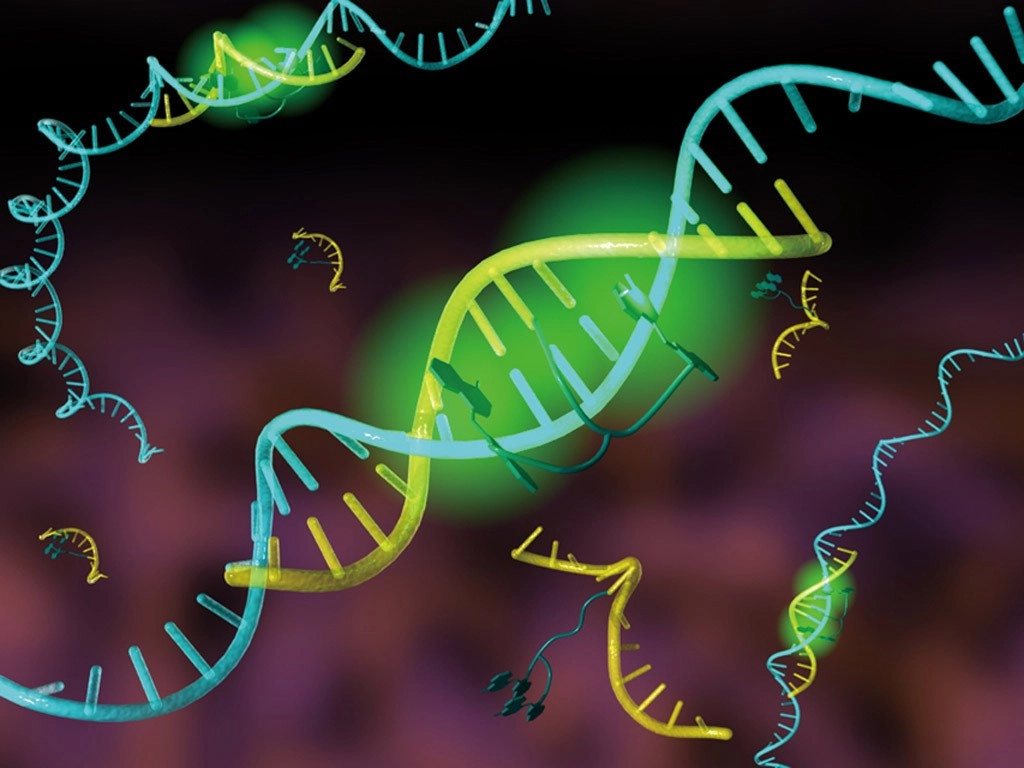
Fluorescence in situ hybridization (FISH) is a powerful molecular technique that allows scientists to visualize the presence and location of specific nucleic acid sequences within a cell or tissue sample. By using fluorescently labeled probes that bind to complementary DNA or RNA sequences, FISH enables researchers to detect and map the position of specific genes, chromosomes, or RNA molecules with high precision. This technique has become indispensable in many fields, including genetics, cancer research, diagnostics, and developmental biology.
Genprice
Scientific Publications

Fluorescence in Situ Hybridization (FISH): A Powerful Tool for Gene and Chromosome Analysis
What is Fluorescence in Situ Hybridization (FISH)?
Fluorescence in situ hybridization (FISH) involves the hybridization of fluorescently labeled nucleic acid probes to their complementary DNA or RNA sequences in a tissue section, cultured cells, or other biological samples. The fluorescent signals emitted by the labeled probes can be observed using a fluorescence microscope, allowing researchers to pinpoint the exact location of specific genetic material in the sample.
FISH can be used to identify specific genes, chromosomes, or regions of interest, making it a valuable tool for mapping the genome and studying gene expression in situ (i.e., within the natural context of the tissue or cell).
How Does FISH Work?
The FISH technique involves several key steps:
- Sample Preparation: The first step in FISH is preparing the biological sample, which may be tissue sections, cultured cells, or blood samples. These samples are fixed to preserve their structure and to prevent degradation of nucleic acids.
- Denaturation: The DNA or RNA in the sample is denatured by heating, which separates the double-stranded DNA into single strands. This step allows the nucleic acid probe to bind to the complementary sequence.
- Hybridization: A fluorescently labeled probe, which is a short segment of DNA or RNA that is complementary to the target sequence, is introduced to the sample. The probe binds to the specific sequence of interest through base pairing.
- Washing: After hybridization, excess or unbound probes are washed away to reduce background noise and ensure that only specifically bound probes remain in the sample.
- Visualization: The sample is examined under a fluorescence microscope. The fluorescent signals emitted by the labeled probes are visualized, allowing the location of the target sequences to be identified.
FISH can be used to detect both DNA (e.g., specific genes or chromosomal regions) and RNA (e.g., mRNA expression in specific cells or tissues).
Types of FISH
There are several variations of the FISH technique, each tailored to specific research needs and applications. Some of the most common types include:
1. Chromosome-Specific FISH (Chromosome Painting)
Chromosome painting uses a mixture of fluorescently labeled probes that are specific to individual chromosomes or chromosomal regions. This method allows researchers to visualize entire chromosomes or specific parts of chromosomes in a sample. Chromosome painting is widely used in cytogenetics to detect chromosomal abnormalities, such as translocations, deletions, or duplications, which are often associated with genetic disorders or cancers.
2. Gene-Specific FISH
Gene-specific FISH targets a particular gene or gene region. This version of FISH is often used to determine the location of specific genes on chromosomes, to investigate gene amplification or deletion, and to examine gene rearrangements in cells or tissues. It is particularly useful in cancer research, where specific gene amplifications or deletions can be linked to the development of certain cancers.
3. Fluorescence in Situ Hybridization for mRNA (RNA FISH)
RNA FISH, also known as RNA in situ hybridization (ISH), uses probes that specifically bind to mRNA molecules in cells. This method is used to detect and localize specific RNA molecules within cells and tissues, providing insights into gene expression patterns. RNA FISH can be used to identify which cells are expressing a particular gene in a tissue, making it an important tool in developmental biology, neuroscience, and cancer research.
4. Comparative Genomic Hybridization (CGH)
Comparative genomic hybridization is a technique that compares the DNA content of two samples, such as a normal and a cancer sample. FISH-based CGH allows researchers to identify chromosomal gains, losses, or amplifications that may be present in one sample but not the other. This method is commonly used in cancer research to study genetic alterations and identify potential therapeutic targets.
5. Single-Cell FISH
Single-cell FISH is a powerful technique used to study gene expression at the individual cell level. This method allows researchers to analyze the expression of specific genes in single cells, providing a more detailed view of the cellular heterogeneity in a tissue or sample. It is particularly useful in stem cell research, developmental biology, and cancer research, where understanding the behavior of individual cells can yield valuable insights into disease mechanisms and treatment strategies.
Applications of FISH
FISH is a versatile technique that has been applied in numerous fields of biology and medicine. Some of the key applications include:
1. Cancer Research and Diagnostics
FISH is widely used in cancer research to detect genetic abnormalities such as gene amplifications, translocations, and deletions, which can be important markers of cancer development and progression. For example, FISH is used to detect the HER2 gene amplification in breast cancer, which is a key diagnostic marker and therapeutic target. Additionally, FISH can be used to monitor chromosomal rearrangements, such as the Philadelphia chromosome in chronic myelogenous leukemia (CML), which helps guide treatment decisions.
2. Genetic Disease Diagnosis
FISH is an essential tool in diagnosing genetic disorders, particularly those caused by chromosomal abnormalities. By using chromosome-specific probes, FISH can detect aneuploidies (e.g., trisomy 21 in Down syndrome), deletions (e.g., DiGeorge syndrome), and other structural chromosomal abnormalities that are often responsible for genetic diseases.
3. Developmental Biology and Gene Expression Studies
RNA FISH is widely used in developmental biology to study gene expression in tissues and embryos. By detecting and localizing mRNA molecules, researchers can track the expression of specific genes during development and identify which genes are active in particular cell types. This provides insights into the molecular processes that regulate development, differentiation, and organ formation.
4. Neuroscience
In neuroscience, FISH is used to study the expression patterns of genes in the brain and other nervous tissues. By visualizing gene expression at the cellular level, researchers can gain a better understanding of how specific genes contribute to neurological diseases, such as Alzheimer’s disease, autism, and schizophrenia. FISH can also be used to map neural circuits and investigate the roles of specific genes in synaptic plasticity and brain function.
5. Infectious Disease Research
FISH can be used to detect specific pathogens in tissues or cells, making it a useful tool in infectious disease research. For example, FISH has been employed to detect bacterial DNA in tissue samples from patients with infections. It is also used in microbiology to study microbial communities in environmental samples or host tissues.
Advantages of FISH
- High Sensitivity and Specificity: FISH is highly sensitive and can detect even low levels of target DNA or RNA. The use of fluorescent probes ensures that only sequences of interest are detected, minimizing background noise.
- Single-Cell Resolution: FISH can be used to analyze gene expression at the single-cell level, providing detailed information about cellular heterogeneity within tissues.
- Visual Mapping: FISH provides a visual representation of gene and chromosome location, allowing researchers to observe spatial patterns of gene expression and chromosomal organization.
- Multiplexing: FISH allows the use of multiple probes simultaneously, enabling the detection of multiple genes or chromosomal regions in a single sample.
Challenges and Limitations
While FISH is a powerful tool, it does have some limitations:
- Resolution: The resolution of FISH is limited by the diffraction limit of light, which can make it difficult to detect very small chromosomal regions or genes in closely packed areas.
- Complexity: FISH requires specialized equipment, including fluorescence microscopes, and can be technically demanding, especially for multiplexing or single-cell applications.
- Probe Design: Designing highly specific probes that bind only to the target sequence can be challenging, and cross-reactivity with similar sequences can lead to false positives.
Conclusion
Fluorescence in situ hybridization (FISH) is a powerful and versatile tool that provides invaluable insights into the structure and function of genes and chromosomes. From cancer research to developmental biology, FISH has enabled scientists to visualize the location and expression of specific genes with high precision, leading to advances in diagnostics, gene therapy, and molecular research. Although it has some limitations, the continued development of FISH techniques promises to enhance its applicability in various scientific and clinical fields.
